Back
Back
|
Respiratory diseases
|
Contagious Pleuropneumonia (Mycoplasma mycoides subspecies mycoides)
Local names:
Luo: Athung'a / Kamba: kyambo / Kipsigis: chebwonit / Embu: kiviti / Gabbra: sombessa / Kikuyu: rimunia / Maasai: olkipei, longishu, ol gibei, ol kibiei / Meru: mohir / Samburu: ikipei / Somali: sambab, harwein, agmar / Swahili: ugonjwa wa mapafu / Turkana: loukoi, lotai / Luvugusu: madjukhu / Nandi: chepuonit / Rendile: ikipei /
Common names:
Peripneumonia, peripneumonie contagieuse (French) peripneumonia contagiosa (Spanish)
Description:
Respiratory
Introduction
| WARNING: Notifiable disease! If you suspect an animal has CBPP or CCPP, you must inform the authorities immediately. |
This is an infectious disease of cattle and goats, caused by bacteria which affect the lungs.
In cattle it is called "Contagious Bovine Pleuropneumonia" (CBPP) and in goats "Contagious Caprine Pleuropneumonia" (CCPP).
Contagious Bovine Pleuropneumonia is widespread in Africa, especially in the semi- arid countries south of the Sahara from West Africa to Somalia. It also occurs in India, China, and South East Asia. Although nowadays mostly confined to the tropics, it is not primarily a tropical disease. In the past it was widespread in Europe, America, Australia and South Africa before being eradicated. In cattle the disease can be acute, subacute or chronic (rapid, mid-term or long term) and is characterised by pneumonia and other lung problems.
Contagious Caprine Pleuropneumonia of goats closely resembles the disease in cattle but is more widespread in Kenya than CBPP. This disease is specific to goats and does not affect sheep, even when goats and sheep are in close contact. It also is a peracute, acute or chronic highly contagious disease characterised by lung problems.
Mode of spread
The disease is transmitted between animals through the inhalation of droplets expelled by infected animals when they cough. The bacteria do not survive for long in the open environment hence direct contact is essential for transmission. Infection spreads faster where the animals are crowded together e.g. in houses, stables, night bomas, markets and during transportation and also in watering points.
The incubation period varies but in cattle most cases occur 3 - 8 weeks after exposure.
A serious aspect of the disease in cattle is the carrier animal, commonly referred to as a "lunger", which appears clinically normal but has the infection in its lungs. The infection is surrounded by fibrous tissue, walling it off from the rest of the lung. It may vary in size from the size of a pea to a large orange, but in time, perhaps after many months, the capsule may break down, allowing the still viable bacteria to escape through the bronchi and infect susceptible animals. The apparently healthy carrier animal therefore, is mainly responsible for the spreading the disease.
"Lungers" do not occur in goats as they do in cattle.
The incubation period is rather less for the disease in goats than in cattle, being 3-5 weeks. The disease in goats is extremely contagious, affecting 100% of goats in a flock and killing 60-100% of them.
The disease in cattle is less dramatic, with lower numbers of a herd affected, sometimes only 10%, although this may rise to 50% with a death rate ranging from 10% to 50% of those infected. Of recovered animals 25% may become carriers with walled?off lung lesions, which cannot be detected.
Signs of Contagious Pleuropneumonia: In Cattle
| Typical stance of cattle affected by CBPP showing extended and arched back, extended head and arched elbows. It has difficulty in breathing and shows "heave marks" from the strain |
- Severe Bovine Pleuropneumonia is characterised by a rise in temperature, accelerated breathing, loss of appetite, and a dejected appearance.
- Later a cough develops, dry at first and then moist. The breathing becomes laboured, accompanied by a painful grunt.
- A characteristic stance is assumed in which the animal faces the wind with its head extended and its elbows turned out in an effort to access more air.
- There may be a nasal discharge containing both mucus and pus and swelling of the lower part of the chest. The animal lies down and after 1-3 weeks it dies. Chronically affected cattle usually exhibit signs of varying intensity for 3-4 weeks, after which the animals appear to recover but they may be dangerous carriers, with walled-off lesions in their lungs.
In Goats:
- In goats there may be sudden death without the appearance of any symptoms.
- In less severe cases of Contagious Caprine Pleuropneumonia there is nasal inflammation and discharge,
- a chronic cough,
- fever up to 41 degrees Celsius ( 106 degrees Fahrenheit),
- weakness, loss of condition, lack of appetite, exercise intolerance, and
- general debility, with death after 2 to 3 days
Diagnosis
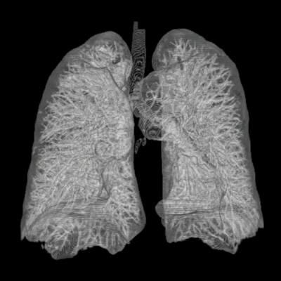
Post mortem lesions are similar in both goats and cattle and are very striking and there should be no difficulty in making a diagnosis. Sometimes only one lung is affected. The lung has a mosaic or marbled appearance. It is no longer soft and filled with air but very firm, solid and dense, requiring force to cut it through with a knife. The lobules, which are consolidated, and gray, yellow or red in colour, are separated from each other by grossly distended septae containing beads of yellow lymph. Straw coloured fluid- up to 10 litres in cattle- fills the chest cavity, with thick, clotted masses of yellow fibrin lying over the exposed lungs. The lungs may be stuck to the chest wall by adhesions.
In goats as much as 500mls of fluid is present in the chest. The lesions are so dramatic and the changes so marked that one wonders how the animal has managed to survive as long as it has.
In Kenya the disease in goats is much more common than that in cattle. In goats diagnosis should not be difficult. The characteristic signs of severe nasal catarrh, respiratory distress, coughing, rapid spread through the flock, the possible recent introduction of goats from another area, the non-transmission to sheep, coupled with the unmistakable post mortem findings should leave no doubt. If there is any doubt samples should be taken for laboratory analysis.
Contagious Bovine Pleuropneumonia occurs less frequently, most often in cattle from the north, which may then transmit to susceptible cattle further south.
Diseases with similar symptoms
Cases in live animals may be difficult to diagnose, being confused with diseases such as ECF (see below) and Pastuerellosis (see below), but a post mortem examination should point the way. Samples of lung should be forwarded for laboratory examination and serum samples taken from in-contact animals.
Prevention - Control - Treatment
Preventive measures
- Contagious Bovine Pleuropneumonia is a Notifiable Disease imposing a strict quarantine on any affected farm, within an endemic area. Animals will be tested and affected animals will be slaughtered. Quarantine may continue for up to 3-4 months after the last clinical case or reactor. Vaccination will not be allowed in non-endemic areas due to the risk of spreading the disease via the attenuated vaccine.
- Vaccination. An attenuated CBPP vaccine injected into the tip of the tail has been used in Kenya in endemic areas. There are developed control programmes in most countries and farmers are encouraged to support, cooperate and work with those programmes. Vaccines against CBPP are used in such control programmes. The control programmes usually start with vaccinating twice per year then once yearly. In developed countries, control programmes usually aim at test and slaughter of all infected animals.
- Sick animals should be isolated and kept strictly separated from healthy animals. The disease spreads by droplet infection so separation is a vital control measure.
- Goats should be routinely vaccinated. The disease is prevalent and regular vaccination using vaccine from KEVAVAPI in Nairobi will do much to lessen the incidence of this devastating disease.
Veterinary assistance should promptly be sought in the control of both of these diseases.
Treatment
Goats may be treated, either with Tylosin Tartrate at a dose rate of 10mg/kg for 3 days or with Oxytetracycline at 15 mg/kg daily, but this should be done at early stages
Treatment of cattle is not justified due to the risk of converting active infection into a carrier state and perpetuating the disease.
East Coast Fever Complex (Theileria parva parva, Theileria parva lawrencii)
Description:
Tick borne protozoal parasite disease.
East Coast Fever is described in detail under Tick Borne Dieseases. Here only a brief summary:
Signs
- The parotid lymph nodes below the ear become enlarged 14 to 16 days after attachment of the tick.
- A few days later a fever develops. This can be up to 41 to 42 degrees Celsius (106 to 107 degrees Fahrenheit).
- Milk yield decreases.
- Other superficial lymph nodes enlarge, especially those in front of the shoulder joint and in front of the stifle joint of the hind leg. These are often clearly visible to the attentive observer.
- Appetite decreases, body condition declines, parasaties appear in the blood and the animal is infective to ticks and hence to other animals.
- There is fluid in the lungs and pneumonia occurs. The animal breathes increasingly rapidly and with increasing distress. It starts to cough, especially if made to move and nasal discharge is common.
- The eyes often appear milky or blue and there may be shedding of tears.
- Sometimes there is diarrhoea which can be violent or blood stained.
- There is muscle wasting and posterior weakness.
- Finally the animal collapses and dies, usually about 18 to 26 days after tick attachment.
Diagnosis
Very often when an animal has died from East Coast Fever or Corridor Disease there is a pile of white froth lying on the ground in front of its nostrils. The sight of this should always stir an observant person into thinking about ECF/Corridor Disease. This froth is due to the massive pulmonary oedema (lung fluid) which has killed the animal.
Post mortem examination will reveal lungs full of fluid and the windpipe full of white froth. Cutting the lungs with a knife will release a flood of fluid from the now clearly defined pulmonary lobules. This is a dramatic, almost diagnostic sign.
Lymph nodes are enlarged and fluid-filled. The kidneys may have white spots on their surface - "infarcts". In the abomasum or fourth stomach may be seen punched out erosions.
A final diagnosis is dependent on identifying the parasite in the animal. Blood smear and gland (biopsy) smears should be taken for laboratory analysis by a qualified vet to confirm the disease.
The overall picture should be taken into consideration. A change in some aspect of management some 2 to 3 weeks earlier, a change of acaricide (tick-killing solution), the appearance of ticks, increased rainfall or improved ground cover leading to extended tick survival, the intrusion of buffalo, illegal cattle movement within the area, all must be considered when formulating a strategy for control.
Apart from the clinical picture, post mortem findings and the examination of blood and lymph node smears there are a number of serological diagnostic tests, but these are of limited value in the field.
Treatment: see here Tick Borne diseases
Pneumonia in young
Calf Influenza, Virus pneumonia, Enzootic pneumonia
Local names
Embu: / Borana & Gabbra: Qufa, Qando/ Luo: / Kipsigis: / Kikuyu: "Rimunia"/ Kamba: Mavui/ Maasai: / Maragoli: / Samburu: Ibus-bus/ Somali: Sombeez/ Turkana: Lotai/
Embu: / Borana & Gabbra: Qufa, Qando/ Luo: / Kipsigis: / Kikuyu: "Rimunia"/ Kamba: Mavui/ Maasai: / Maragoli: / Samburu: Ibus-bus/ Somali: Sombeez/ Turkana: Lotai/
Description
Management disease
Management disease
Introduction
Pneumonia is a very common disease of calves/lambs/kids and is an important cause of losses before weaning. Surviving animals often show reduced growth and may not be worth keeping.
Causes
It is atypical multi-factor disease. Infectious agents take advantage of the contributing factors to make the animal sick. Very important contributing factors are:
- Wet and cold condition
- Crowding and stress
- General weakness because of poor nutrition
- Following other infections that weaken the animal (e.g. chronic diarrhea)
- Weak born calves
- Lack of colostrum
Some of the infectious agents are:
- Viruses: Respiratory syncytial virus, Para-influenza type 3, Infectious bovine rhinotracheitis IBR and many others
- Bacteria: Streptococci, Pasteurella, Chlamydia (esp. In kids/lambs)
- Aspiration pneumonia due to forced feeding of milk or forced drenching with drugs; liquid gets into the lungs
Signs:
- Rise in temperature, which can be as high as 40.5 - 42 degress Celsius accompanied by watery discharge from the eyes and noses. Rapid breathing and coughing. High fever can cause trembling/shivering.
- Later the cough becomes dry and hard and the discharge becomes thick (pus).
- More severely affected calves stand with their heads down, backs arched and breath very heavily with an effort to get enough air
- Calves become very dull and are unable to stand up, the coat becomes rough. This is a very bad sign and such calves are likely to die.
- In acute cases of pneumonia there is also sudden death.
- Aspiration pneumonia: animal coughs immediately after forced feeding/drenching, develops severe breathing problems and can die within two days
Prevention - Control - Treatment
Prevention and Control
Proper ventilation to minimize draughts in a calf house
- Avoid over crowding of calves in the house (best are single calf pens) and pasture
- Provide dry, warm bedding to prevent chilling and ensure good ventilation This will also help to lower the overall humidity in side the calf pen.
- Avoid mixing calves of different age groups and especially calves from different sources.
- Isolate sick calves from the rest if possible
- Calves must receive fresh colostrum at 8-10% of body weight during first 3 hours after birth but not later than 12 hr after birth.
- Aspiration pneumonia: avoid forced feeding, only experienced people should drench young animals
Treatment
The best chances for treatment with antibiotic and Sulphonamide is when the disease is still fresh. Calves with difficult breathing should also be given anti-inflammatory drugs in addition to antibiotic treatment. Normally calves respond within 3 days but treatment should be continued for around five days.
Good selection of drugs for treating pneumonia is:
Good selection of drugs for treating pneumonia is:
- Penicillin-Streptomycin combination and other penicillin
- Tetracycline
Start with one antibiotic first and observe improvement. If fresh cases do not respond to treatment consult a veterinarian.
Tuberculosis (Mycobacterium bovis)
Local names:
Luo: Kahera / Kiswahili: Kifua kikuu / Turkana: Lokud / Maragoli: Kehera / Maasai: Enkirhoget / Samburu: nkiroget /
Luo: Kahera / Kiswahili: Kifua kikuu / Turkana: Lokud / Maragoli: Kehera / Maasai: Enkirhoget / Samburu: nkiroget /
Common names:
pearls disease, phthisis, consumption, tuberculose (French)
pearls disease, phthisis, consumption, tuberculose (French)
Description:
Zoonotic disease
Zoonotic disease
Introduction
| WARNING: Notifiable disease! If you suspect an animal has Tuberculosis, you must inform the authorities immediately. |
Tuberculosis is a bacterial infection that affects mainly cattle and human beings, as well as other domestic animals. Mycobacterium bovis is the usual cause of tuberculosis in cattle, while M.avium causes the disease in poultry and M. tuberculosis is responsible for the majority of cases in man. It occurs worldwide but prevalence is dependant on the type of husbandry and the efficacy of control measures. Its prevalence is low in cattle kept extensively and not housed at night.
| WARNING: Consumption of raw milk by humans should be discouraged. People get tuberculosis from animals. Never drink unboiled milk. |
Mode of spread
The bacterium is transmitted when an infected animal excretes the organism in various discharges such as exhaled air, saliva, nasal discharges, faeces, urine and milk. The germs can survive outside the host for several weeks as long as they are not exposed to harsh climatic conditions like direct sunlight.
Housed animals become infected when they inhale the germs and grazing animals can be infected through feed supplementation and water troughs., Calves may be infected by drinking milk from infected cows.
In some countries wildlife are involved in transmission of tuberculosis to cattle. Badgers in Britain, brush-tailed possums in New Zealand,white-tailed deer, elk and bison in North America, and and feral pigs in Australia are cases in point.
Tuberculosis is usually a chronic disease, that is, it may last several months during which affected animals lose condition and may eventually die.
Signs of Tuberculosis
- In the early stages there are no outward signs. In cattle, the infection starts with the formation of a lesion in the lung. The infection then spreads to the local drainage lymph node.
- If the mycobacteria overcome the host's defenses, lesions in the organs and lymph nodes will progressively enlarge. Abscesses will form which contain thick pus which may even calcify.
- Bacteria may perodically escape to infect adjacent tissues or organs, or pass via the blood or lymph to other organs.
- If in the lungs a bronchopneumonia may develop. Single very large abscesses may occcur, or many smaller ones affecting a wide variety of organs.
- Cattle with lesions in the lungs develop a persistent soft cough, decreased appetite, lethargy, weakness, a low-grade fluctuating fever, and progressive loss of weight.
- Later there may be impaired breathing, followed by an increased breathing rate and and discharge of yellowish secretion from the nose.
- Superficial lymph nodes may become enlarged.
- The udder, if infected, becomes enlarged, firm and nodular.
- Lesions in the intestine or other internal organs lead to gradual loss of condition.
Diagnosis
- Diagnosis on clinical signs alone is very difficult. Enlargement of peripheral lymph nodes may be suggestive but no more than that.
- In tuberculous mastitis the signs often involve inflammation found at the base of the quarter of the udder with painless swellings. In tuberculous metritis the signs include yellow coloured pus.
- Accurate diagnosis of tuberculosis requires the application of the Tuberculin Test. It is injected by a vet into the skin on the side of the animals neck and the reaction is read after 72 hours. The presence of any swelling means the animal is infected. This test in infected cattle is highly sensitive. Sensitivity develops within 4 weeks of infecton - so recently infected animals may therefore pass the test.
- Animals in advanced stages of tuberculosis may not respond to the test but such cases are usually emaciated and should be detectable by clinical examination.
- In areas where infections due to M.avium, M.tuberculosis or Johne's Disease occur, causing false positive reactions, a Comparative Test is used. Avian tuberculin is innoculated at one site and bovine tuberculin at a second site. A bovine reaction larger than the avian indicates M.bovis infection.
Diseases with similar symptoms
Any wasting disease such as malnutrition, parasitism, John's disease, CBPP, Traumatic reticulo-pericarditis, mineral deficiencies, sleeping sickness and lung abcesses
Prevention
- Keep cattle premises clean and hygienic at all times. Clean feed troughs, water troughs and disinfect cattle premises.
- Milk fed to calves should be sterilized.
- Sick animals should be isolated.
- Sick people should not handle animals. People infected with tuberculosis caused by M. bovis can transmit infection to cattle.
- Enclosing and, in particular, housing animals aids transmision through air and leads to high infection rates.
- The stress of repeated pregnancies and lactations weakens host defences, allowing the disease to develop progressively, until it becomes clinically signficant. Thus, tuberculosis is seen most often in housed dairy cattle, especially older anmals.and where hygiene is poor
- Mycobacteria may survive on shaded pasture for weeks or months and in yards or houses. Direct sunshine rapidly kills the organisms.
- Never sell or buy sick animals
Treatment and Control
Treatment and vaccination play no part in the control of tuberculosis in animals. Both appear to be ineffective in practice.
Ideally control of bovine tuberculosis is test and slaughter or test and segregate. The former relies on the slaughter of animals that test positive, with repeat testing every 3 months.
Fortunately the disease is not nearly so prevalent in Kenya as it is in more temperate countries where animals are housed for several months of the year due to cold weather during the winter months.
No traditional treatment recommended.
- Ask your veterinary officer or veterinary surgeon for advice and assistance
Review Process
Created by:
1. Draft by: William Ayako Aug 2009
2. Updating and editing Dr Hugh Cran April 2010
3. Review workshop team. Nov 2 - 5, 2010:
1. Draft by: William Ayako Aug 2009
2. Updating and editing Dr Hugh Cran April 2010
3. Review workshop team. Nov 2 - 5, 2010:
- For Infonet: Anne, Dr Hugh Cran
- For KARI: Dr Mario Younan KARI/KASAL, William Ayako - Animal scientist, KARI Naivasha
- For DVS: Dr Josphat Muema - Dvo Isiolo, Dr Charity Nguyo - Kabete Extension Division, Mr Patrick Muthui - Senior Livestock Health Assistant Isiolo, Ms Emmah Njeri Njoroge - Senior Livestock Health Assistant Machakos
- Pastoralists: Dr Ezra Saitoti Kotonto - Private practitioner, Abdi Gollo H.O.D. Segera Ranch
- Farmers: Benson Chege Kuria and Francis Maina Gilgil and John Mutisya Machakos
- Language and format: Carol Gachiengo
Information Source Links
- Barber, J., Wood, D.J. (1976) Livestock management for East Africa: Edwar Arnold (Publishers) Ltd 25 Hill Street London WIX 8LL
- Blood, D.C., Radostits, O.M. and Henderson, J.A. (1983) Veterinary Medicine - A textbook of the Diseases of Cattle, Sheep, Goats and Horses. Sixth Edition - Bailliere Tindall London. ISBN: 0702012866
- Blowey, R.W. (1986). A Veterinary book for dairy farmers: Farming press limited Wharfedale road, Ipswich, Suffolk IPI 4LG
- Force, B. (1999). Where there is no Vet. CTA, Wageningen, The Netherlands. ISBN 978-0333-58899-4.
- Hall, H.T.B. (1985). Diseases and parasites of Livestock in the tropics. Second Edition. Longman Group UK. ISBN 0582775140
- Hunter, A. (1996). Animal health: General principles. Volume 1 (Tropical Agriculturalist) - Macmillan Education Press. ISBN: 0333612027
- Hunter, A. (1996). Animal health: Specific Diseases. Volume 2 (Tropical Agriculturalist) - Macmillan Education Press. ISBN:0-333-57360-9
- ITDG and IIRR (1996). Ethnoveterinary medicine in Kenya: A field manual of traditional animal health care practices. Intermediate Technology Development Group and International Institute of Rural Reconstruction, Nairobi, Kenya. ISBN 9966-9606-2-7.
- Merck Veterinary Manual 9th Edition
- Pagot, J. (1992). Animal Production in the Tropics and Subtropics. MacMillan Education Limited London
- Sewell & Brocklesby. Handbook on Animal Diseases in the Tropics 4th Edition
- The Organic Farmer magazine No. 50 July 2009
- The Organic Farmer magazine No. 51 August 2009