Back
Back
|
Diagnosis
|
| Introduction | Description of Symptoms | |||
| Diagnosing Live animals | Review Process | |||
| Post mortem | Information Source Links | |||
| Taking Samples |
Introduction
When animals get sick farmers most often need the services of the veterinarian to help find out what is wrong with their animal as livestock are expensive and keeping them healthy is a priority.
However in East Africa there is a severe lack of skilled veterinarians and farmers often accept the loss of animals without a chance of knowing why the loss came about. There are in Kenya several veterinary investigation laboratories and often veterinary clinics at a distance. Farmers can do a lot to find the cause of a disease and get it diagnosed by telephone and/or by delivering in samples for analysis.
There are several outward signs that can help in identifying the cause of the problem, but often they are not sufficient to determine with a good degree of certainty what the exact nature of the disease is. And treating an animal for the wrong disease is not only expensive and useless, it also gives the animal additional stress.
The following is an attempt to guide non-vets in examining sick or dead animals to find the cause of the problem.
Diagnosis is when:
- The vet helps finding the cause of disease by examing symptoms of sick animals and possibly blood and lymph node samples.
- The vet (or skilled farmer) carries out a post mortem (opening up the animal after death to find where the problem lies).
- The vet takes various samples and either examines them under a microscope or sends them to a laboratory to get the causative agent of disease identified. For example when there is an outbreak of Foot and Mouth Disease it is very important to identify the strain of the virus so that correct vaccination can be carried out in non affected animals in the neighborhood to prevent further spread. This can only be done in a good veterinary laboratory.
- The vet uses all the results from the different methods of analysis along with symptoms before death to arrive at the correct diagnosis and recommends a suitable treatment where applicaple.
Diagnosing Live animals
Hopefully the animal is still alive and you only need to call the vet and describe the symptoms and get recommendations for treatment. The vet may ask you if you are able to take smear samples of blood and lymph nodes and bring in. This is a simple procedure that can easily be learned - if you want to learn please see below under Taking Samples. Please now take pen and paper and note down which of the following conditions you see on a piece of paper before you call the vet:
1. Appearance of the animal
a) Expression:- anxious, feverish, nervous, excitable, aggressive, dull, lethargic
b) General condition:- weak, thin, emaciated, dropsical
c) Skin:- weeping, dry, peeling, scurfy, crusted, lumpy, bald, bearing lice, ticks, stick-tight fleas,
d) Coat:- matted, dry, rough, staring.
e) Mucous membranes:- (the linings of the eyelids and natural openings):red, blueish, yellow, pale; with vesicles, with pustules, ulcers (sores), haemorrhages (collections of blood), cheesy deposits, necrotic areas, smelling badly.
f) Eyes:- cloudy, discharging, protruding, sunken, bloodshot
g) Lymph nodes:- enlarged, normal
h) Temperature: High, low, subnormal, fluctuating. A rectal thermometer is very useful to any livestock keeper.
i) Behaviour such as Coughing, shivering, dilated nostrils, facing the wind, groaning, grunting, grinding the teeth, salivating, looking at the flank, rolling, convulsions, staring, staggering, blindness, turning in circles, star-gazing, high-stepping, vomiting, arching the back, avoiding light, failing to chew the cud, paralysis, coma.
2. Natural functions
a. Appetite and thirst:- reduced, increased, capricious, depraved
b. Respiration:- shallow, quick, slow, difficult, laboured, irregular
c. Heartbeat:- slow, fast, thumping, irregular, with pulse in jugular vein
d. Defaecation and urination:- frequent, suppressed, painful, forcible
e. Faeces:- hard, slimy, soft, frothy, clay-coloured, black, greenish, containing blood clots, shreds of mucous membranes, worms
f. Urine:- pale, yellow, blood-tinged, viscid, cloudy, strong-smelling, forming a deposit on standing, containing blood clots
g. Milk:- yellow, pink, with white clots, blood clots, ropy, smelling
3. Discharges
Continuous or intermittent, clear, cloudy or containing pus, watery or ropy, yellow, pink, blood-streaked, foul-smelling, from what source and how much of it is there.
4. Swellings
Hot or cold, hard or soft, painful or painless, containing liquid or gas, pitting or crackling on pressure, tense or flabby, sharply or ill-defined, discharging, pointing ( soft at one part), how distributed and of what size.
Post mortem
Post mortem is the name of an examination after death carried out to determine the cause of death. Any animal which dies without an obvious cause, whether after a long or short illness, should be subjected to a post mortem examination. Not to do so leaves the cause of death in the realms of speculation and supposition. But by doing a post mortem much may be learned both to the benefit of the farmer and to the other animals in the flock or herd.
The post mortem should always be carried out by a qualified veterinarian who is trained in determining even slight changes from normality in animal tissue. However where no vet is available and samples have to be transported to a laboratory, the following procedure can be followed.
Tools Required
The basics needed are a sharp knife, scalpel, panga or axe, rubber gloves, note pad and pen, glass slides, swabs, leakproof plastic bags or pots, protective clothing, gum boots and overalls. A digital camera is very useful to record observations which may be difficult to put into words.
It should be borne in mind that certain diseases of animals are transmissible to humans e.g. Rift Valley Fever, Q Fever, brucellosis, salmonellosis, rabies, anthrax and salmonellosis. Therefore questions should always be put to the owner regarding the history of the animal about to be post-mortemed.
If an animal is found dead without obvious cause, then before the carcase is opened a check should be made to ascertain whether it had died from anthrax. This is done by making a blood smear from a drop of blood taken by nicking the ear, spreading it on a slide, staining it and examining it under a microscope. Such examination should be done by a qualified veterinarian. Remember that Anthrax is a Notifiable Disease and is easily transferred to humans, so be very careful not to touch any blood with bare hands.
Pregnant women should NOT carry out post-mortems, especially those of sheep, as they run the risk of having a miscarriage from infections such as those caused by toxoplasmosis, Q fever etc.
Carcases to be post-mortemed should be as fresh as possible in order to gain the best results.
Adequate light and plenty of water are essential. A raised working area is helpful.
Procedure
1. Start with a thorough external examination. Note the general body condition and make the following notes or dictate so someone taking notes while the post mortem is going on, and if there is a camera nearby it is good to also take pictures:
- Is the animal fat, thin, emaciated?
- Is there any discharge from the nose, eyes, ears, vulva or rectum?
- Is there any evidence of diarrhoea?
- Open the vulva and look at the conjunctiva to check on the colour? Is it normal, pale, yellow, red?
- When did the animal die?
- Is it bloated?
- Look at the eyes. Are they clear and moist, or opaque and blue? Sunken or protruding? Discharging or bloodshot?
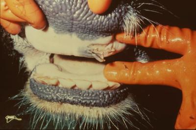
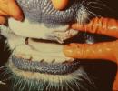
- Open the mouth and examine the tongue and gums. Note any ulceration, erosions or redness. Look at the nostrils, checking for any discharge or erosions.
2. Next carefully remove the skin, noting whether it peels easily or with difficulty perhaps indicating dehydration. Are there any haemorrhages (blood collections) below the skin or any oedema (swelling)?
3. Place the carcase on its back and carefully cut though the midline, taking care not to cut into the rumen if it is bloated. Cut through the sternum (middle of chest) with an axe or panga to open the chest cavity. Check for any abnormal fluids in the chest or abdomen such as pus or serous fluids.
Before removing any organs look at how they are situated in the chest or abdomen and make notes.
Before removing any organs look at how they are situated in the chest or abdomen and make notes.
- Are the lungs lying freely, or do they stick to the chest wall?
- What colour are they? Are they a normal pink or are they excessively pale or are they dark red or even purple?
Cut along the length of the neck and dissect out the windpipe, gullet and tongue and reflect them backwards and remove them together with the lungs and heart. Cut the diaphragm where it joins the chest wall and dissect backwards to remove the stomach, liver, spleen, kidneys and intestines.
Lay everything out on a smooth, clean, washable surface and examine in a logical manner.
4. Begin at the front end and work backwards. And always make notes.
- Open the windpipe. Is there any froth of fluid present? Examine the lymph nodes in front of the shoulder. Are they enlarged and moist, or small and dry?
- Examine the lungs. Are they pink and light or dark and heavy? Do they sink in water or do they float? Cut into the lungs. Does any fluid flow out? Do you see any froth? Is any air trapped within the lung tissue?
- Examine the heart. Open the pericardial sac (Bag around the heart). There should be a small amount of fluid between it and the heart. Are there any visible adhesions (does the heart stick to the surrounding bag?)Open the heart. Is the blood inside the heart clotted or unclotted? Any foreign bodies such as a wire should be visible.
- Is the spleen soft and enlarged, or small, shrunken and dry?
- Check the liver. Check its colour. Is it liver-coloured or yellowish? Cut across it, noting its consistency and that of the bile ducts. In chronic liver fluke disease the bile ducts may be gritty and calcified. Is the liver sticking to the diaphragm (the wall between stomach and lung/heart region)?
- Check the gall bladder for size and the appearance of its contents.
- Check the urinary bladder. Open it carefully and note the colour of the urine within, assuming that it is still full. The urine may be red, brown, yellow or clear.
- Next examine both kidneys for size and consistency. Cut them length wise and note what you see and the colour and form of any urine within.
- Open the rumen (first stomach) and empty out the contents. Are there any foreign bodies present e.g. nails, wire, plastic bags etc? Do you see any unusual plant material? Examine the rumen surface for parasites such as rumen flukes, which may also be found in the reticulum. If present note their number and position.
- Examine the reticulum for foreign bodies as this is where they most frequently lodge.
- Cut into the omasum and note the consistence of its contents. Are they dry or moist?
- Next open the abomasum, or 4th stomach, taking due note of the consistency of its contents. Examine the surface of the abomasum. Is it pale and swollen, or reddened and ulcerated? This is where worms, such as Haemonchus contortus, will be found in lambs, adult sheep and calves. Write everything down or dictate to someone who will.
- Open the small intestine, noting the colour and consistency and smell of its contents. Is the surface pale or reddened and inflamed? Are the contents fluid or dark and sticky? Is there any blood present? Examine closely for the presence of parasites such as intestinal worms.
- Proceed to the large intestine, again noting the consistency, colour and smell of the contents. There may be constipation in diseases such as Anaplasmosis.
5. Now proceed to examine the abdominal (stomach related) organs.
6. Now proceed to the gastro-intestinal tract. Open the gullet from the throat to the rumen and note what you see.
7. Finally examine the musculature for any swelling, discolouration, redness or blackening, such as might occur in certain clostridial diseases such as blackquarter.
Taking Samples
A full account of the post-mortem findings, in conjunction with a description of any symptoms prior to death is very important to reach a trustworthy diagnosis.
To cut down on volume for taking to the lab the most important samples are those that appear not to be normal.
|
|
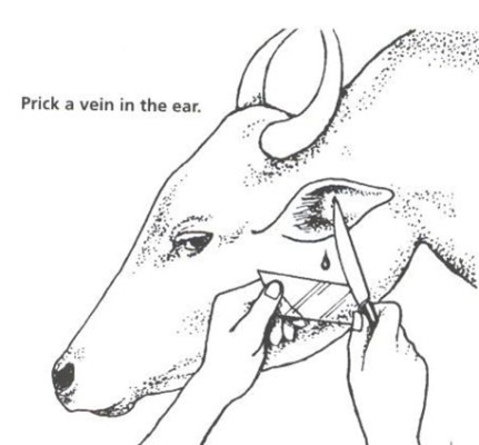
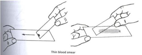
1. Blood smears should be taken. Disinfect a small area at the base of the animal's ear. Prick a small hole with a disinfected needle so that a drop of blood appears. A small drop of blood is then touched with a clean glass slide. With another glass slide touch the drop with one end of the slide until the blood spreads along the angle between the two slides. Then push the upper slide along the length of the lower slide so that it draws the blood after it. Wave the slide in the air and it should dry almost immediately if the smear is of the correct degree of thinness. Place inside a clean envelope. Do NOT stick two smears together. So doing renders both useless for diagnostic purposes.
2. Take a gland smear by inserting an 18 gauge needle attached to a 2 ml syringe into a prescapular (those in front of the shoulder blade) lymph node , suck back on the syringe and expel the sucked up fluid onto a clean slide.
3. Fresh faecal samples should be put into clean polythene bags and be sealed and labeled.
Other samples MUST be presented FRESH to a veterinary surgeon or veterinary officer. Bringing in a bag of decomposing, toxic, smelly organs will not advance the diagnosis, but it will certainly make you very unpopular.
Description of Symptoms
If the animal was also seen alive before death then a good clinical description of the symptoms together with a clear PM report will all help the contacted veterinarian to reach a diagnosis, in conjunction with submitted smears, faeces and possible fresh material.
If the list of symptoms while the animal was still alive is considered while any patient is examined it may draw attention to points which might otherwise be overlooked, so please before you submit the report to the veterinarian also include the symptoms before death noted down as described above.
A clearly written post-mortem report, together with a brief but straightforward description of any symptoms which occurred prior to death, plus blood and lymph node smears and fresh samples if possible will go a long way towards helping the consulted veterinarian to reach a diagnosis.
Information Source Links
- John Gilmour 1992: Making the Most of Ovine Necropsy. In Practice Journal of Veterinary Postgraduate Clinical Study Volume 14 No 3 May ISSN No 0263/841 X
- MJ Fitzpatrick 1950: Notes on Animal Diseases, The Diagnosis of Animal Diseases in the Veterinary Laboratory. Veterinary Research Laboratories, Veterinary Department Kabete, Kenya. Originally printed in the East African Agricultural Journal