
 Back
Back
|
Calf problems
|
| Introduction | Coccidiosis | |||
| Calf White scours (calf enteritis) | Review Process: | |||
| Pneumonia | Information Source Links | |||
| Salmoneliosis (calf paratyphoid) |
Introduction
Many diseases of newborn calves can be controlled with proper hygiene and health management. Although calves will inevitably be exposed, minimizing risk factors will result in fewer infections and illness in calves.
Calf White scours (calf enteritis)
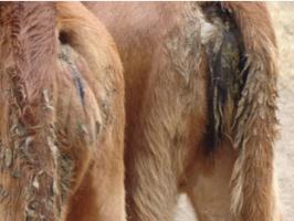 |
| Dirty calf tails typical sign of calf scours |
| © William Ayako, Kari Naivasha |
Common names: Calf enteritis
Description: Management disease
Description: Management disease
Calf white scoursis is a disease in which bacteria present in the intestinal tract become active and disease causing. It is common in calves up to a few months old and is mainly regarded as a disease of bad management. It is encouraged by overcrowding of calves in pens, damp housing caused by poor ventilation and bad feeding particularly abrupt over feeding with milk.
Signs of calf white scours
- Bad smelling diarrhea which is usually whitish in color and sometimes frothy. In older calves, the color of the dung is darker.
- The affected calves lack appetite, are dull and listless.
- The calf develops fever at first signs and loses weight very fas
Prevention and Control
- Better hygiene in the calf house is a good control measure.
- It is important to reduce the amount of milk from the first day of the infection. Instead give 2 bottles of warm water mixed with two table-spoonfuls of glucose three times a day then add whole milk gradually mixed with warm water from the second day. Resume whole milk feeding after the calf has fully recovered.
- Immediate separation of the affected calves from the rest of the calves.
- Provision of warm dry beddings and correct feeding regime
- Regular administration of calf scour serum to calves in farms where the disease is endemic is important.
Recommended treatment
Use of sulphonamide drugs and antibiotics. given on time normally give good results.
Pneumonia
Common names:
Calf Influenza, Virus pneumonia, Enzootic pneumonia
Description: Pneumonia is the most common of all the diseases of weaned calves and is the main cause of highest losses in weaned calves in terms of mortality and reduced growth rates. It is often associated with scours, weakness or loss of condition caused by scours, poor feeding or bad housing. The disease usually ends in death, and recovered animals may not be worth keeping.
Description: Pneumonia is the most common of all the diseases of weaned calves and is the main cause of highest losses in weaned calves in terms of mortality and reduced growth rates. It is often associated with scours, weakness or loss of condition caused by scours, poor feeding or bad housing. The disease usually ends in death, and recovered animals may not be worth keeping.
Some of the causative agents include:
- Viruses e.g. the Respiratory syncytial virus, Para-influenza type 3, Infectious bovine rhinotracheitis, Bovcine viral diarrhea and Coronavirus
- Bacteria e.g. Pasteurella multocida and Pateurella haemolytia, Haemophilus somnus and the Corynebacterium pyogenes.
- Mycoplasmas e.g. Mycoplasma dispar, Mycoplasma bovis and the Acholeplasma laidlawii
Signs of Pneumonia
- Rise in temperature which can be as high as 40.5 - 42 ºC accompanied by watery discharge from the eyes and noses.
- Rapid breathing and cough
- More severely affected calves stand with their heads down, backs arched and breath very heavily with an effort to get enough air.
- In acute cases of pneumonia there is sudden death
Diagnosis
The disease can be diagnosed through post mortem examination of the fresh lungs and in the living animal through the history and symptoms.
Prevention and Control
- Proper ventilation to minimize draughts in a calf house
- Avoid over crowding of calves in the house and pasture
- Provide dry, warm bedding to prevent chilling and to reduce ammonia and other noxious gases which damage the normal protective mechanisms in the calf's respiratory system. This would also help to lower the overall house humidity.
- Avoid mixing calves of different age groups and especially calves from different sources in the same air space.
- Maintain good levels of antibody protection by giving adequate colostrums and by vaccination.
Recommended treatment
Sulphonamide and antibiotic treatment are very effective when given in good time. However, prolonged use of long acting penicillin and tetracycline injection given twice weekly for three weeks or more is good for chronic lung infections.
Salmoneliosis (calf paratyphoid)
Common names:
Calf paratyphoid
Description: Bacterial infection
Description: Bacterial infection
Salmoneliosis, also called calf paratyphoid, is a bacterial infection which affects all animals including cattle, birds, pigs, sheep and humans. It is more serious in young calves and weaned calves and can cause a wide range of symptoms.
In young milk fed calves, the disease is caused by a serotype Salmonella called Salmonella typhimurium. In weaned calves, the main cause of the disease is Salmonella Dublin.
The infection is carried from symptomless carriers, which is either the dam or other weaned calves when groups are mixed at weaning.
It can also be transmitted through contact with contaminated pastures and feeds, housing, and contaminated stagnant waters.
Infection occurs after ingestion of causative agent. The affected herd may remain infected with Salmonella dublin for several years without causing a disease
.In young milk fed calves, the disease is caused by a serotype Salmonella called Salmonella typhimurium. In weaned calves, the main cause of the disease is Salmonella Dublin.
The infection is carried from symptomless carriers, which is either the dam or other weaned calves when groups are mixed at weaning.
It can also be transmitted through contact with contaminated pastures and feeds, housing, and contaminated stagnant waters.
Infection occurs after ingestion of causative agent. The affected herd may remain infected with Salmonella dublin for several years without causing a disease
Signs of Salmoneliosis
- In young calves when the disease is caused by S.typhimurium, the characteristic symptoms include the following:
- In severe form, there is profuse yellow diarrhea with a very bad smell accompanied with septicemia and high temperature. Death may occur within 24 hours or less.
- In chronic form, the affected calf would have pasty dung, and is often unthrifty. In some cases, some calves would carry the disease without suffering adverse effects. This group of calves is called intermittent excretors. They may sometimes shed salmonella in their feces when they become stressed thus affecting other calves.
- In weaned calves, affected with S.dublin,symptoms include:
scouring which resemble the one caused by S.typhimurium may occur.
Septicemia, pneumonia, or meningitis.
Sometimes, scouring may be seen containing lumps of intestinal wall, blood and mucus. Infected calves would be dull, lack appetite and may also be coughing.
Occasionally, a group of calves may appear thrifty abruptly and sudden deaths may be observed.
Prevention and Control
- Isolate the sick calves to reduce the likelihood of infection to the remainder of the group. This is important because some infected animals do not show signs when they have the disease.
- Avoid feeding feeds contaminated with feces to calves.
- Vaccination using a live vaccine is a good control measure and this can be given from day old or soon after purchase of the calf.
- Give adequate colostrums (as soon as possible and as much as possible) to boost immunity of the calf soon after birth.
- Gentle treatment of purchased calves, e.g. good warm bedding, separate penning, feeding of fluid therapies and gradual increase of milk.
- Avoid purchasing replacement calves instead, it is advisable to rear your own replacement calves.
Recommended treatment
It is advisable to treat the animals as soon as possible with recommended antibiotics. The same drugs can be administered through the mouth or by injection. Coccidiosis
Common names:
Description: Parasite infection
Description: Parasite infection
Coccidiosis is a disease affecting all animals but it has serious negative economic consequences in young animals. The disease is caused by a group of protozoan parasites known as eimeria. This group of protozoa does not have a carrier like others such as the one causing malaria in humans or trypanosomiasis in cattle.
The eimeria protozoa are believed to be present in adult animals and can therefore be picked by young animals from water. The Eimeria zurnii is particularly responsible for causing Coccidiosis. It burrows in the wall of the lower gut, thus causing the disease. Incubation takes about 3 weeks from the time of infection. Weaned calves and young stock between the ages of 2 months and 2 years are susceptible to the infection.
Signs of Coccidiosis
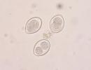
- The affected calves pass out semi solid feces, usually with variable quantities of chocolate-brown blood mixed with it. The most characteristic clinical sign of coccidiosis in calves is straining to pass out blood stained feces technically known as tenesmus.
- The calves would appear dull, weak and lose weight rapidly
- They display discomfort and grind their teeth
Diagnosis
Faeces from the infected animals should be taken to a veterinary laboratory for microscopic examination.
Prevention and Control
- Ploughing and reseeding of infected grazing pastures would help to minimize the disease occurrence in areas where the disease is prevalent
- Careful rotation of calves in the grazing paddocks helps to minimize infection
- Avoid confining calves to one grazing area for a prolonged period
- Rest grazing paddocks for an ample time to avoid accumulation of pathogens
Recommended treatment
Immediate administration of anticoccidiaosis therapy such as sulphonamides, nitrofurazone and amprolium by injection or by mouth
Review Process:
1 Draft By William Ayako, Animal scientist, KARI Naivasha Aug 2009
2 Review workshop team. Nov 2 - 5, 2010
2 Review workshop team. Nov 2 - 5, 2010
- For Infonet: Anne Bruntse, Dr Hugh Cran
- For KARI: Dr Mario Younan KARI/KASAL, William Ayako - Animal scientist, KARI Naivasha
- For DVS: Dr Josphat Muema - Dvo Isiolo, Dr Charity Nguyo - Kabete Extension Division, Mr Patrick Muthui - Senior Livestock Health Assistant Isiolo, Ms Emmah Njeri Njoroge - Senior Livestock Health Assistant Machakos
- Pastoralists: Dr Ezra Saitoti Kotonto - Private practitioner, Abdi Gollo H.O.D. Segera Ranch
- Farmers: Benson Chege Kuria and Francis Maina Gilgil and John Mutisya Machakos
Information Source Links
- Barber, J., Wood, D.J. (1976) Livestock management for East Africa: Edwar Arnold (Publishers) Ltd 25 Hill Street London WIX 8LL. ISBN: 071310063X
- Blood, D.C., Radostits, O.M. and Henderson, J.A. (1983) Veterinary Medicine - A textbook of the Diseases of Cattle, Sheep, Goats and Horses. Sixth Edition - Bailliere Tindall London. ISBN: 0702012866
- Blowey, R.W. (1986). A Veterinary book for dairy farmers: Farming press limited Wharfedale road, Ipswich, Suffolk IPI 4LG
- Force, B. (1999). Where there is no Vet. CTA, Wageningen, The Netherlands. ISBN 978-0333-58899-4.
- Hall, H.T.B. (1985). Diseases and parasites of Livestock in the tropics. Second Edition. Longman Group UK. ISBN 0582775140
- Hunter, A. (1996). Animal health: General principles. Volume 1 (Tropical Agriculturalist) - Macmillan Education Press. ISBN: 0333612027
- Hunter, A. (1996). Animal health: Specific Diseases. Volume 2 (Tropical Agriculturalist) - Macmillan Education Press. ISBN:0-333-57360-9
- ITDG and IIRR (1996). Ethnoveterinary medicine in Kenya: A field manual of traditional animal health care practices. Intermediate Technology Development Group and International Institute of Rural Reconstruction, Nairobi, Kenya. ISBN 9966-9606-2-7.
- Pagot, J. (1992). Animal Production in the Tropics and Subtropics. MacMillan Education Limited London. ISBN 0-333-53818-8
- The Organic Farmer magazine No. 50 July 2009
