Back
Back
|
Abortion and Stillbirth
|
Introduction
As is described below, there are many more causes of abortion than brucellosis. The inadequacy of current labs in the country/region to diagnose causes of abortion different from brucellosis and possibly Campylobacter/Trichomonas, is a major constraint on identifying them. However close observation and a gathering of a full history should go some way towards reaching a tentative diagnosis.
- Abortion is the termination of pregnancy after the development of the foetus is complete, but before it can survive.
- Stillbirth is the expulsion of a dead full-term foetus, whose lungs were not inflated.
- Early embryonic death is when pregnancy ends before the full development of the foetus.
An attempt to diagnose every abortion is rarely justifiable economically, given the low diagnostic success rate, the high cost involved, and the poor profit margin in the beef and dairy sectors. However if the foetal loss is more than 3-5% per month or year then there should be concern and efforts made to identify the cause.
|
DANGER: Some causes of abortion are highly infectious. When handling animal tissue after an abortion make sure you wear plastic gloves and do not touch any of the tissues with bare hands. All tissue and samples to be take to a laboratory must be safely stored in sealed plastic bags with no leakage.
All other abortion/stillbirth tissue must be disposed of by burning or deep burying to protect from scavengers
|
Diagnosis
Diagnosis of any cases of abortion is a complex process and one which requires skilled professional help. Therefore in such instances consult your veterinarian.
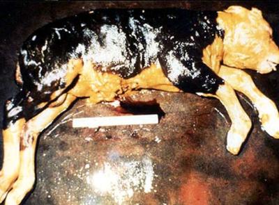
Diagnosis of abortion in livestock can be a difficult and frequently frustrating business. The diagnostic success rate is pretty low - 30-40% for cattle and 60-65% for sheep. This is due to several reasons. Often abortion follows the initial infection after weeks or months by which time the causal agent is no longer apparent. An aborted foetus may be so decomposed that no diagnosis is possible. Often the aborted foetus and its placenta are contaminated by environmental agents. Non infectious causes of abortion, such as toxic or genetic influences may be of such a nature that easy diagnosis is not possible.
To get the best possible specimen for analysis it is recommended to take the entire foetus and placenta, together with a serum sample from the mother. The placenta and foetus should be cleaned with water or saline, packed in clean plastic bags, chilled but NOT frozen, and rapidly transported to the nearest laboratory.
The decomposition rate in the foetus is much slower than in the carcase of an animal which has been born alive, so if chilled quickly, most foetuses will be suitable for examination, even if they do not reach the lab for 1-2 days. With sheep and goats bring the entire foetus and placenta. If this is not possible, then collect stomach or abomasal contents, heart blood or fluid from a body cavity, lung, liver, kidney and spleen and a serum sample from the mother. Try to submit in sterile containers. Because they are always contaminated do not mix placenta with other tissues.
The reason for submitting stomach contents is that most abortion causing agents, especially bacteria and fungi, infect the placenta and then gain entry from the stomach of the mother into the amniotic fluid which is swallowed by the foetus.
A single antibody sample in the mother rarely provides evidence of abortion caused by a particular agent. Often abortion occurs weeks or months after the initial infection and by the time abortion occurs the disease causing agent is stable or declining. If the herd in general is infected, paired serum samples taken 2 weeks apart from 10% of the herd, or a minimum of 10 animals, often demonstrate seroconversion and provide evidence of active infection in the herd.
Non-infectious Causes of Abortion
A good history and physical examination of the aborting cow (or ewe) are particularly important for conditions in this category, as in most cases diagnosis in unlikely to be achieved by laboratory examinations alone.
Physical
Abortion may be caused by trauma (shock/sickness), hyperthermia due to maternal fever, insemination, and twinning. High environmental temperatures are not likely to cause abortion. The foetus is well protected by its surrounding of amniotic fluid so incidents like being pressed in a crush are not likely to cause abortion.
Nutritional
Some factors may lead to primary or secondary deficiency such as those caused by:
- Iodine deficiency has been associated with stillbirths and weak newborn calves. This may due to lack of Iodine as occurs in certain areas of Kenya .
- Feeding of kale to livestock.
- Application of human sewage sludge to pasture.
- Selenium/Vitamin E deficiency has been associated with bovine abortions as has.
- Vitamin A deficiency.
Toxic (poisons)
Various toxins, such as those which occur in a number of plants, may cause abortion. These may act by producing excessive amounts of pre-formed oestrogens , which, by acting on the corpus luteum , result in abortion. Cattle frequently abort following an outbreak of nitrate/nitrite poisoning (after heavy fertilization of pastures with nitrogen fertilizer) which poisons the dam and subsequently also in the foetus. Common sources of nitrate/nitrite on farms are fertilisers and certain plants.
Signs of poisoning
Clinical signs of poisoning include:
- severe difficulty in breathing,
- muscle twitching and
- brown mucous membranes and blood are likely to be present.
Genetic
Genetic abnormalities may result in congenital malformations causing foetal death and consequent abortion. Bulldog calves are one example. Early abortion or early embryonic death may also occur.
Infectious Causes of Abortion.
These may be divided into abortion caused by bacteria, viruses, protozoa and fungi.
Other severe general infections of the dam, such as Anaplasmosis, East Coast Fever, and acute mastitis may also lead to foetal death and abortion.
Bacterial Causes
Brucella abortus
The abortion rate of Brucella abortus may be up to 90% in susceptible herds.
For more information on Brucellosis click here. In some countries Brucellosis has been eradicated or brought under control by a programe of blood testing, slaughter and heifer vaccination. However in most parts of Kenya/East Africa there has been no control or vaccination and brucellosis is common in both humans and animals.
Brucellosis in humans resemble malaria, and is often mistaken as such unless identified through laboratory testing. Cure of brucellosis in humans is a very long drawn out and expensive administration of antibiotics and much better avoided by controlling the disease in livestock.
Cattle/sheep/goats/pigs and camels/donkeys become infected by eating the organism from abortion material, genital discharges, or contaminated pasture, food or water. Abortion occurs 2 weeks to 5 months after the initial infection and generally occurs after the 6th month of pregnancy. There is an endometritis, placentitis and abortion. Retained foetal membranes are common signs. Calves if born alive are weak. In non-pregnant animals the udder and supra-mammary lymph nodes are colonized and infection of the placenta may occur during the next gestation.
Brucellosis is often present in the raw milk milk, which presents a serious risk to human health as this is a dangerous zoonosis. Always boil milk before consumption!
Diagnosis is by maternal serology or isolation of the organism from placenta, foetus or uterine discharge. (For more information please link to Brucellosis).
The aborted placenta is often oedematous (swollen) and leathery, with necrosis (wilting) of the cotyledons.
Prevention of Brucellosis
A single vaccination of all female animals between 5 and 8 months of age with S19 vaccine will give adequate immunity for the lifetime of the livestock. Adult vaccination should be avoided if possible to allow serum sampling in an outbreak of abortions to be interpreted without the confusion caused by antibodies due to vaccination with the live S19 vaccine.
Salmonella species
For further information see here
In many countries Salmonella dublin is endemic in cattle and is the most common species of Salmonella causing abortion. Many other Salmonella species may be involved in individual cases. Aborting cows often have a fever and diarrhoea, although some may show no symptoms other than abortion.
All species of Salmonella may infect man and cause food poisoning so are called zoonotic.
Outbreaks may be spasmodic or may affect several members of a herd.
Abortion may occur at any time, but often occurs between 6 and 9 months.
No specific lesions are seen in the placenta or the foetus.
Leptospirosis
Up to 30% of a herd may abort with an outbreak of Leptospirosis. The disease is zoonotic and food poisoning by Leptospirosis in humans may be mild, severe or fatal (causing death).
Leptospirosis is an important zoonosis, with urine and milk being infected for up to 3 months. Humans are susceptible to all types found in domestic animals. Contact with surface water contaminated by urine from infected carcases, or contact with the tissues from infected animals is the most common source of infection.) For further information see here
Sorurces of infection:
- The infection may be transmitted venereally
- Via urine and abortion material.
- Watercourses and standing and stagnant water can become contaminated and be a source of infection.
- Sheep and goats may be "silent" carriers and a potential source of infection.
- Rodents may also act as carriers and be a source of infection.
Outbreaks are more common when the weather is wet and there is standing water to hold infective organisms.
Symptoms of Leptospirosis
Cattle may abort without symptoms of illness, or may be acutely ill with severe jaundice (yellowing of gums and white of eyes), fever, haemoglobinuria, photosensitzation (for further information see here), and death.
The aborted placenta is avascular with yellow-brown cotyledons and oedematous (swollen), yellowish inter-cotyledonary areas.
The foetus usually dies 1-2 days before expulsion and so is decomposed.
Prevention by:
- Vaccination with a multi-valent vaccine, every 6 months is recommended where Leptospirosis is prevalent.
- Sources of infection, such as contaminated water and feed should be identified and eliminated.
- Rodents and other vermin should be destroyed. It should be noted that cattle may be life-long carriers of leptospirosis and remain a source of infection.
Leptospira interrogans, serovar hardjo, grippotyphosa, pomona, canicola, and icterhaemorragiae may cause abortions in the last third of pregnancy, 4-6 weeks after maternal infection.
Diagnosis from Laboratory:
is by detection of leptospires in foetal kidney, culture and examination of maternal urine, and serological testing of maternal blood samples.
Campylobacter (Vibrio)
For further information see here
The organism Camplobacter fetus fetus and venerealis cause early embryonic death, infertility and occasionally abortion. This is a venereal disease and most outbreaks occur after the introduction of an infected bull into a susceptible berd.
Most cases of abortion, when they occur, are between 4 - 8 months of pregnancy.
Usually there are no significant findings other than a mild inflammation of the placenta.
Diagnosis is by identification and culture of isolated organisms or response to a vaginal mucous agglutination test, or by examination of preputial sheath washings. The organism is very fragile, making laboratory diagnosis a very frustrating and in many cases an impossible goal.
Control is by :
- Vaccination and the use of artificial insemination.
- The infected bull may be treated with parentral Streptomycin, together with oil-based Streptomycin applied to the penis. It is not usually practicable to treat infected cows, which generally recover spontaneously.
Listeriosis
Listeriosis is a zoonosis so may spread to people and cause food poisoning.
The organism Listeria monocytogenes is widespread in the environment being present in the soil, sewage effluent, bedding and feedstuffs. Animals get the disease by eating infected feedstuff. The infection then travels to the placenta, causing inflammation.
A substantial number of outbreaks are associated with the feeding of silage, especially if the silage contains soil.
Signs of Listeriosis:
- Encephalitis and eye infections, are rarely seen in conjunction with abortion.
- The foetus is usually decomposed when aborted.
- There is a placentitis, with multiple, yellow or gray necrotic spots in the cotyledons and similar spots in the liver of the aborted foetus.
Abortion usually occurs in late gestation and is usually sporadic, although rarely abortion storms may occur.
There is no vaccine. Control should involve an awareness of the risks of feeding silage contaminated by soil.
Bacillus licheniformis
This organism is frequently associated with sporadic abortion in cows and is widespread being especially common in silage, feedstuffs and bedding contaminated with silage effluent and wet spoilt hay. The organism probably spreads following eating infected feedstuffs, then travels via the bloodstream to the placenta, causing late abortion.
Abortions occur in late pregnancy and the placenta is dry and leathery and yellowish-brown.
Arcanobacter pyogenes
This organism may cause sporadic abortion at any stage of pregnancy. It is present in the noser ans throat of many normal cows and in abscesses. When it gains entry to the bloodstream it causes an endometritis and placentitis. There is no vaccine.
Viral Causes
Bovine Viral Diarrhoea
For further information see here: BVD/Mucosal Disease
This is the most commonly diagnosed virus in bovine abortion cases.
The pathology of BVD in the developing foetus is complex.
Infection before 125 days results in foetal resabsorption, muumification, or expulsion.
Foetuses that survive infection with non-cytopathic BVD virus at between 80 - 125 days gestation develop immumotolerance to the virus and subsequently become persistently infected, but frequently die between 6 - 24 months of age.
Foetal infection at between 100 and 150 days gestation often results in the development of congenital defect.
Late gestation infection between 125 - 285 days is cleared by the foetus as it is now immunocompetent.
The persistently infected animal acts as a potent source of infection for susceptible animals. Those persistently infected animals that do not die of Mucosal Disease may live long enough to join the adult herd and produce persistently infected calves.
Diagnosis is on maternal blood - a more than 4 fold increase in antibody titre in paired serum samples obtained 2 weeks apart verifies recent infection. Virus may be isolated from blood, nasal swab specimens or from other tissues. Other tissues for virus isolation include spleen, lymph nodes and ulcerated segments of intestine.
Outbreaks of abortion by organisms that normally cause sporadic abortion should raise the suspicion of possible concurrent BVD infection.
Prevention focuses on the removal of infected animals and herd vaccination.
Vaccines are modified live virus vaccines and inactivated viral vaccines. The latter should be used in pregnant cattle.
Infectious Bovine Rhinotracheitis
This disease is caused by a herpesvirus and usually results in respiratory disease, but may also cause abortion in cows up to 100 days after infection. Abortions generally occur in late pregnancy and aborting cows may or may not have shown respiratory signs previously. When there is a fever resulting from systemic or respiratory disease this can cause abortion.
Diagnosis is difficult due to the expulsion of the foetus before it is infected, which is usually decomposed.
Abortion is generally sporadic but can be up to 60% of susceptible animals in the herd.
Cows may be carriers of the virus.
Control is by herd vaccination and intranasal, modified live virus and killed vaccines are available.
Rift Valley Fever
For further information see here
One of the main symptoms of this epidemic viral infection is abortion in several species, including cattle and camels. The epidemic nature of this disease should help to identify its nature. It is also a zoonosis affecting man, both directly and indirectly.
Fungal Causes
Fungal placentitis is an important cause of sporadic abortion.
Abortion occurs from mid to late pregnancy.
Abortion occurs from mid to late pregnancy.
Mouldy hay, straw, silage and brewer's grains are important sources of infection. Among the most commonly isolated fungi are Aspergillus and Mucor species. Fungi are inhaled and travel via the bloodstream to the placenta. Gray ringworm-like lesions may be seen on the head and shoulders of the foetus, which is not usually decomposed although it may be dehydrated.
Diagnosis is based on isolation of fungi from the foetal stomach, placenta and skin lesions.
Control is based on avoidance of mouldy feed.
Protozoal Causes
Neospora caninum
This organism of which the dog is the definitive host occurs worldwide and is a common cause of abortion in some countries and occurs in Kenya.
Abortion most commonly occurs between 4 and 6 months of gestation. Sporadic abortions or abortion storms may occur and repeat abortions in affected cows have been reported. Congenital transmission with vertical infection of calves in utero occurs with recrudescence of disease in chronically infected cows. Transmission from cow to cow does not occur. Occasionally infected calves are born alive with paralysis.
Usually the aborted foetus is decomposed, cows are not clinically ill and the placanta is not retained.
If infected calves are born alive they remain infected for years and possibly for life. Then during pregnancy organisms may become activated and infect the foetus. This is thought to be the most common source of infection.
There is no treatment but a commercial vaccine is available. ( Not in Kenya !)
Strict hygiene to prevent faecal contamination of feed by dogs may aid in prevention. Strictly keep dogs out of feeding areas.
Trichomonas fetus
This organism is transmitted venereally and usually causes infertility and early embryonic death, but may also cause abortion in the first half of pregnancy.
The placenta is often retained and there may be pyometra - pus in the uterus. The foetus has no specific lesions although T. fetus may be found in abomasal contents, placental fluids and uterine discharges.
Vaginal mucous swabs and sheath washings may be used in diagnosis to retrieve the organism.
Control is by using artificial insemination using non-infected bulls.
Virgin bulls may be used in heifers.
Virgin bulls may be used in heifers.
Abortion in Sheep
| Warning: Pregnant women should NOT deal with lambing ewes or any aborted material, and persons, whether pregnant or not, should be aware that many agents which cause abortion in sheep, are transmissible to humans. e.g. Toxoplasma, QFever, Salmonella, Leptospira and Listeria. |
Abortion in sheep, as in cattle, is not always easy to diagnose.
As in cattle abortion may occur weeks or months after an infection has been acquired and often there is little that can be done to halt an outbreak once it is in progress. However, there is still a need for investigation in order to make a diagnosis from material submitted at the time so that management decisions can be made for future years, based on the confirmed cause.
A detailed history is essential, giving details of numbers of ewes, expected lambing dates, feeding, vaccinations, numbers of abortions, and date.
Aborted fetuses and placentas should be submitted for examination.
Ewes which have aborted should be isolated, aborted products should be disposed of by burning or deep burying and protection from scavengers, and if the ewe is ill, or has retained her placenta, antibiotic therapy should be initiated.
Do not foster lambs onto ewes which have aborted.
Some causes of abortion are similar to those which cause abortion in cattle. e.g. Salmonellosis, Listeriosis, Neospora caninum, Brucellosis and Leptospirosis. Others have been detailed in separate data sheets, e.g. Q Fever and Toxoplasmosis - for further information see here
There are some causes of abortion which target sheep specifically, and these include EnzooticAbortion of Ewes and Border Disease.
Diagnosis of any cases of abortion is a complex process and one which requires skilled professional help. Therefore in such instances consult your veterinarian.
Enzootic Abortion of Ewes
This is caused by a bacterium called Chlamydophila abortus, which can also infect people.
Infection is via mouth and is not apparent until the next pregnancy, when the organism invades the placenta at ninety days gestation, coinciding with the phase of rapid foetal growth.
If rams are infected, they may develop inflammation of the testicles and the organism is excreted in the semen, although venereal spread is most unlikely.
Signs of Enzooic Abortion
- Abortion storms, involving up to 30% of ewes in a flock, are a feature of EAE, especially in intensively managed flocks. Ewes are not noticeably ill. They have solid immunity after an abortion.
- Abortions occur in the last three weeks of pregnancy and weak, live lambs may be born alongside dead lambs.
- The placenta between the cotyledons is thickened with a brownish exudate over the surface. The foetuses are usually fresh.
The organism is present in large numbers in the abortion products and vaginal fluids for up to three weeks, leading to contamination of the environment and infection of other sheep. Some ewes become carriers.
The organism is present in large numbers in the abortion products and vaginal fluids for up to three weeks, leading to contamination of the environment and infection of other sheep. Some ewes become carriers.
Diagnosis
Examination of fresh placenta is the method of choice for the diagnosis of abortion due to C. Abortus. Alternatively, vaginal swabs or swabs taken from the coat of aborted fetuses can be used.
Prevention and Control
Various vaccines are available, including an inactivated vaccine for use in pregnant animals. They are not available in Kenya .
Whole flock treatment with long-acting oxytetracycline can be used 3 - 6 weeks before lambing, to increase the number of viable lambs born to ewes known to be infected with C. abortus.
In the face of an outbreak, mark and isolate aborting ewes, dispose of bedding and abortion products and disinfect pens. Ewes that have aborted should not be used to foster lambs.
Border Disease
This is caused by a virus related to the one that causes Bovine Viral Diarrhoea/Mucosal disease in cattle. For more information see here BVD/Mucosal disease
The virus usually enters a flock through the purchase of a persistently infected replacement. Problems are seen at the next lambing, i.e. barren ewes, stillbirths, abortions and persistently infected lambs which will continue to act as a source of infection for the rest of the flock. P.I. females that reach breeding age may have reduced fertility and will either abort or produce live, P.I. lambs.
Signs of border disease
As with BVD, the outcome depends on the stage of gestation at which infection takes place. In non-pregnant sheep infection is usually inapparent, leading to long-lived immunity.
- In early pregnancy (less than 60 days gestation), infection may lead to foetal death with subsequent resorption, mummification, abortion or stillbirth. A proportion will survive as persistently virus affected lambs. These are the so-called "hairy shakers", with coarse wool over the head and neck, tremors and doming of the cranium, and an abnormal appendicular skeleton. Poor growth rate is also apparent.
- Between 60 and 85 days gestation, the outcome is unpredictable. Lambs are usually strongly antibody positive, but may show neurological signs.
- Infection after 85 days may lead to abortion, but most lambs will survive with antibodies to BDV.
Diagnosis
This is on samples of blood and brain tissue.
Prevention and Control
Prevemntion is achieved by operating a closed flock system and testing all replacements. As there is no local laboratory capable of testing for presence border disease, any system here must depend on close observation. If "hairy shaker" lambs are born in large numbers, then segregate in-lamb ewes from those with affected lambs and, as infection during pregnancy results in an immunity to re-infection, mix susceptible animals retained for breeding with surviving hairy shakers through the grazing period to maximize their chances of becoming immune.
There is no vaccine.
There is no vaccine.
Review Process
Information Source Links
- In Practice Journal of Veterinary Postgraduate Clinical Study Volume 29 No 8 September 2007 Abortion in Cattle: causes and investigation Emma Cabell ISSN no 0263/841 X
- In Practice Journal of Veterinary Postgraduate Clinical Study Volume 12 no 3 May 1990 ISSN No 0263/841 X Diagnosing the cause of bovine abortion Geoff Pritchard
- The Merck Veterinary Manual 9th Edition 2005 ISBN Number 0-911910-50-6
- Diseases of Sheep Edited by W B Martin 1983 ISBN 0- 632- 01008-8
- Veterinary Medicine A Textbook of the Diseases of Cattle, Sheep, Pigs, Goats and Horses Blood, Radostits and Hen