Back
Back

|
Insect Borne Diseases: Anaplasmosis, East Cost Fever, Babesiosis, Rift Valley Fever
|
Anaplasmosis
Introduction
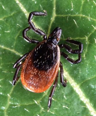
Anaplasmosis an acute febrile disease of cattle, usually transmitted by blue ticks and caused by a rickettsial bacterium, called Anaplasma marginale. This is an obligate intracellular parasite which multiplies by binary fission in red blood cells, causing a severe anaemia. There are other species of Anaplasma. Severe anaplasmosis is caused by Anaplasma marginale. This parasite occurs at the edge of the red cells. A mild,usually inapparent form is caused by Anaplasma centrale. This parasite occurs at the centre of the red blood cell.
The incubation period of the disease is about 2 - 12 weeks and is directly related to the infective dose.
The disease occurs throughout the tropical and subtropical regions of the world
Anaplasmosis is not contagious.Transmission does not take place by contact but generally via the medium of an infected tick vector such as the blue tick.This is a one-host tick, spending its entire life on its host and for whose control once-weekly dipping or spraying is generally appropriate. The source of infection is always the blood of an infected animal. Biting flies, contaminated instruments, injection needles, and oxpecker birds can also transmit infecton. Wild animals and other domestic animals can be infected and become reservoirs of infection.
Infections in the sucking young are usually clinically inapparent, and, once infected, animals remain carriers for life. In areas where cattle first become infected with Anaplasma marginale early in life, losses due to anaplasmosis are minimal. In animals less than 1 year old anaplasmosis is usually subclinical, in yearlings and 2 year olds it is moderately severe, and in older cattle it is severe and often fatal.
Thus exposure of calves to infected ticks confers upon them an often life-long resistance. Paradoxically efficient tick control at this stage of bovine life will later expose cattle to possible life-threatening anaplasmosis. These facts must be kept in in mind at all times.
Zebu cattle, with their relative resistance to heavy tick infestations, are less likely to be
infected, but they are just as susceptible as European breeds.
Carrier animals serve as reservoirs for further transmissions.
Serious losses can occur when mature cattle with no previous exposure are moved into endemic areas or when transmission rates are insufficient to ensure that all cattle are infected before reaching the more susceptible adult age. This latter situation can occur when a preiously effective acaricde loses its efficacy, especially when that efficacy has continued from calfhood into adulthood.
Signs of Anaplasmosis
Clinical signs
Onset of illness is characterised by a rising fever of up to 41 C ( 106 F) , a drop in milk production, and a decreased appette. Acutely affected animals lose condition rapidly and peracutely affected animals may die within a few hours of the onset of clinical signs. Acutely affected animals are depressed, lose their co-ordination, if exerted, become breathless, and lag behind the rest of the herd. If forced to walk some of these animals may even lie down. These symptoms are directly related to the degree of anaemia caused by the destruction of red blood cells. Examination of the eyes and vulva will reveal a change from a healthy pink to a pallid white to a yellow or even light orange colour, indicating the onset of jaundice due to liver damage. The teats of a milking animal appear pale or white in colour.
There is a rapid bounding heart rate. This is easily heard by pressing one's ear against the animal's pelvis. This loud beat, heard several feet from the heart itself, is almost disgnostioc for Anaplasmosis. It cannot be heard in a normal animal.
The urine may be yellow or even brown, but, unlike Babesiosis (Redwater) contains no red blood cells.
Constipation is common.
Pregnant animals often abort.
Some animals are hyperexitable and aggressive, charging and attacking people. In the severe form of the disease, there may be death if immediate treatment is not given. In dead animals, blood is thin and watery and the flesh is pale yellow. The liver is yellowish orange. The gall bladder is large and full of brownish greenish fluid and the kidney is large and soft. The spleen is enlarged and mushy.
Diagnosis
A presumptive diagnosis can be made, based on an assessment of the history, clinical and post mortem signs, together with the appearace of ticks and an anaemia without haemogolobinuria. A peripheral blood smear from the ear should be taken for laboratory analysis by qualified personnel to confirm the disease.
Anaplasmosis is generally not such a severe disease as Babesiosis (Redwater) with which it might be confused. In the latter the urine contains red blood cells, sometimes is almost black in colour and the individual death rate is usually much higher.
Anaplasmosis is generally not such a severe disease as Babesiosis (Redwater) with which it might be confused. In the latter the urine contains red blood cells, sometimes is almost black in colour and the individual death rate is usually much higher.
Prevention - Control - Treatment
Preventive measures
Preventive measures
Regular dipping and spraying with effective acaricides to control tick infestations are vital preventive measures. In non-East Coast Fever areas, and in most extensive systems, young calves are often not dipped for the first few months.This allows them to contract the disease and confers immunity. This will provide an enzootic stability in association with a sub-clinical infection.
The drugs of choice for treatment are tetracyclines and imidocarb dipropionate-Imizol.
- Oxytetracycline long acting solution is effective given early in the disease at a rate of 30mg per kg i/m. Likewise imidocarb at 3mg/kg. These dosages will not elimiate the carrier state.For this repeated injections at a higher dose level are required, which may not be appropriate in Kenya at the present, due to the widespread abundance of the organism in recovered, immune animals.
- Constipated cattle should be given a dose of Epsom Salts - 500g (1lb) is a suitable dose for an adult animal. Careful nursing and provision of an appropriate diet with green feed will assist recovery.
- Eradication is generally not practicable due to the ubiquity of the carrier ticks, the long period of infectivity in carrier animals, the presence of carriers in the wild animal population and the difficulty of identifying infected animals.
- Vaccines are available and used in some countries. These are either livng attenuated strains of A. Marginale coupled with treatment if required, or the less pathogenic A.Centrale. Severe reactions can occur with both types of vaccine.
East Coast Fever
Introduction
The disease is also referred to as Theileriosis. It is a serious febrile disease of cattle transmitted by the brown ear tick and the red legged tick. The infective tick Rhipicephalus appendiculatus usually attaches to the ear and thus called brown ear tick. The disease occurs in east and central Africa. Other forms of the disease are widespread in tropical and subtropical regions of Africa, Asia and southern Europe.
It is caused by a protozoan parasite Theileria parva which invades the lymphatic glands. Incubation varies between 1-3 weeks after exposure to infective ticks.
|
Signs of East Coast Fever
In the event of infection, the parotid lymphnodes below the ear become enlarged 1 - 2 weeks after infection. A few days later, fever develops along with enlargement of superficial lymphnodes in front of shoulder and stifle.
Other signs may include: difficult breathing, soft cough due to accumulation of fluid in the lungs, blood stained diarrhea, muscle wasting and white discoloration of the yes and gums. Cows may appear disturbed resulting in the so called "turning sickness" and paralysis.
Diagnosis Blood smear and gland (biopsy) smears should be taken for laboratory analysis by a qualified vet to confirm the disease.
Prevention - Control - Treatment
Prevention and Control Regular dipping of cattle should be maintained. However, this may be difficult to attain in open herds without proper organization. Treatment o clinical signs as they appear in extensive herds.
The use of live vaccines of East Coast Fever is effective but may cause clinical disease.
Infection and treatment with antibiotics such as long acting oxytetracycline provides immunity.
Recommended treatment The following treatments should be administered by trained personnel:
|
|
Babesiosis
Introduction
The other names of this disease include Red water and Cattle tick fever. This is a febrile disease of cattle, caused in Africa by the intraerythrocytic protozoan parasites Babesia bigemina and Babesia bovis. In Kenya the one host Blue tick, Boophilus decoloratus, which is the main transmitter, can only transmit B. Bigemina and as a result this species assumes more importance than B. bovis. The disease occurs worldwide but has serious implications in the tropics because of the ubiquity of the host ticks.
The distribution of bovine babesiosis caused by B.bovis and B.bigemina is confined strictly to the distribution of their vector tick. This is particularly marked in Africa, to which the tick which transmits B. bovis, Boophilus microplus (Cattle tick ) was introduced at the end of the 19th century. B.bovis thus only occurs in those areas of Africa to which Boophilus microplus has spread, because the indigenous Blue tick only transmits B.bigemina.
Tranmission occurs tranovarially. In endemic areas two features are important in determining the risk of clinical disease.
1) Calves have a degree of immunity (related both to colostral-derived antibodies and to age) that persists for about 6 months.2) Animals that recover from Babesia infections are generally immune for life. Thus, at high levels of tick transmisson, all newborn calves will be infected by Babesia by 6 months of age, show few, if any, clinical signs and subsequently be immune. This situation of enzootic stability can be upset by either natural (eg. climatic) or artificial (eg. acaricide treatment) reduction in tick numbers to levels such that tick transmission of Babesia to calves is insufficient to ensure all are infected during this early critial period.
On recovery from initial infection cattle are immune but harbour the parasite (premune) and may remain carriers for up to 2 years. In enzootic areas, constant reinfection ensures that such cattle remain carriers permanently, although the premunity may break down due to other factors such as stress or intercurrent disease, resulting in clinical babesiosis. Calves, as previously mentioned, are less susceptible than adults and, if infected in calfhood, signs may be minimal.
Thus in enzootic areas two epidemiologial states are recognised:
1) Enzootic stablity:
The continuous presence of Babesia in both cattle and ticks with frequent acquisition of the parasite from active infective ticks ensures that cattle are infected in calfhood and remain carriers due to contant reinfection. Under these conditions clinical babesiosis is minimal.
2) Enzootc instability:
The infection of Babesia is such that not all cattle are infected as calves, and therefore some animals remain susceptible. If these are infected as adults, they may suffer severe clinical reactions. Such conditions arise when the number of ticks fluctuate due to factors such as seasonal variation, drought, dipping etc.
The incubation period ( the time from tick attachment to the appearance of parasites in the red blood cells) is normally in the range of 8 -16 days for B.bovis but at least 9 days longer for B.bigemia, because infection is not transmitted until the nymph and adult stages of the tick begin to feed. The shorter incubation period for B. bovis is due to tranmission by the earlier larval stage of the tick.
Ticks acquire babesia infections from infected animals.
Ticks acquire babesia infections from infected animals.
Signs of Babesiosis
Babesiosis or Redwater is a very severe disease, considerably more so than Anaplasmosis, and the first indication that the disease is present may be the finding of a dead animal - usually an adult - in the paddock.
The parasites invade the red blood cells where they multiply and break out to invade more red blood cells. The released parasite and host constituents from the destroyed red cells are toxic, resulting in various physiological disturbances and shock. The destruction of the red cells causes anaemia and tissue anoxia. The release of haemoglobin results in jaundice and haemoglobinuria - blood in the urine. Sometimes the urine is a dark red in colour. Sometimes it can be almost black. Such animals are very ill indeed and usually die.
There is a rise in temperature. This is often 41C ( 106F) or higher. Affected animals become depressed, listless, rumination ceases, milk yield drops, and they refuse to eat. Pregnant animals often abort. The eyes and gums are pale from anaemia and jaundiced, due to bile pigments in the blood stream.
Occasional animals infected wth B.bigemina show cererbal babesiosis manifested by incoordination followed by posterior paralysis,or by mania,convulsions and coma. Such animals rarely recover, despite treatment.
In less severe cases of the disease, infected cattle have fever lasting about 1 week and remain sick for 3 weeks. This group of animals may recover slowly while pregnant animals may abort. Recovered animals remain permanent carriers of the disease.
Zebu cattle appear to have more resistance to babesiosis than do European breeds.
Post mortem fndings reveal an enlarged and friable spleen, a swollen liver, with an enlarged gall bladder containing dark green granular bile, congested dark-coloured kidneys, and generalised anaemia and jaundice. The urine is usually, but not always, red in colour.
A presumptive diagnosis can be confirmed by the examination of a stained blood smear taken from the ear or tail tip. B.bigemina parasites are large organisms, seen within the red cells in pairs at an acute angle to each other. B.bovis are small and lie at an obtuse angle to each other
A number of serological tests are available such as the indirect fluorescent antibody test, ELISA, and the complement fixation test but generally these are out of reach under local conditions.
A fever, associated with blood in the urine, anaemia and jaundice, with the finding of Babesia organisms in the red blood cells, is diagnostic.
Other diseases must be considered when an animal is seen to be passing red urine. Possibly the most important of these is Leptospirosis. This disease also presents with red urine, jaundice, abortion, depression and loss of appetite. However there are no blood cells in the urine, it affects all ages, including young calves, the degree of jaundice is often more profound due to the more chronic nature of the disease, photosensitisation of white skin is common and it often occurs during periods of heavy rain as rodents frequently are carriers, passing the infection in their urine, which can then survive for extended periods of time in pools of water.
Heartwater may be initially confused with cerebral babesiosis but there are clinical differences and no babesia parasites will be found in stained smears.
Anaplasmosis is a less severe disease with no blood cells in the urine.
Prevention - Control - Treatment
Prevention and Control
At the moment in Kenya prevention must rely on tick control and trying to reach a situation of enzootic stablity by aiming to control the ticks but not the disesase they transmit so that young stock are infected and develop a lifelong immunity. This can be difficult in areas where other ticks transmitting other diseases such as East Coast Fever occur. The Blue tick is normally the first tick to develop resistance to new acaricides. The results of this resistance can be catastrophic.
It is advisable to keep suitable breeds of animals in disease endemic areas. Such breeds may include indigenous cattle and their crosses with exotic cattle. With these breeds the dipping or spraying intervals can be extended as these local breeds have more resistance to babesiosis and also to ticks. But with European breeds tick protection is required. The whole picture must be considered before taking any decision regarding extension or reduction of dipping or spraying intervals.
Vaccination using live, attenuated strains of the parasite has been used successfully in a number of countries. Combined vaccinaton and strict control of ticks is an effective control strategy when exotic cattle are introduced. Young animals can be vaccinated with live vaccines to counter periods of inactivity of Boophilus vectors. These techniques are currently unavailable in Kenya so a carefully monitored tick control program is required to allow the development of resistance in conjunction with the control of other tick-borne infections.
Recommended treatment
Two drugs are currently available for the treatment of babesiois.
These are Diminazene aceturate (Norotryp, Berenil etc) and Imidocarb dipropionate ( Imizol)
Dimiazene is given at a dose rate of 2 to 5 mg/kg by intramuscular injection.
Imidocarb is given at a rate of 1.2mg/kg by subcutaneous injection.
Higher doses of both drugs will protect animals for several weeks.
Large species of Babesia such as Bigemina are more responsive to treatment than the small species such as Bovis which may require a larger dose of the same drug to effect a cure. Fortunately Bigemina is the more common species in Kenya.
The aim of treatment is to promote recovery from the clinical disease while allowing some organisms to persist in order to allow the animal to develop an immunity to the disease. Too rapid a cure can result in the animal being susceptible to reinfection.Thus, treatment which immediately sterilizes the infection should, if possible,be avoided. This can be difficult when faced with severe cases when delayed treatment means death of the animal. In such cases, prophylactic treatment of the rest of herd should be considered.
Supportive treatment is advisable. This may include the use of non-steroidal anti-inflammatory drugs, multivitamins, fluid replacement and in some cases, blood transfusions.
Rift Valley Fever
Introduction
Rift Valley Fever is an acute, or peracute, mosquito- borne viral disease affecting domestic ruminants - cattle, sheep, goats, camels and domestic buffaloes- and man. It occurs mainly in East and Southern Africa and more recently in Saudi Arabia and Yemen. During epidemics the occurrence of numerous abortions, deaths in young animals (and adults) - and acute symptoms in humans tends to be characteristic.
The virus is widely distributed in Africa, but major epidemic episodes in animals and humans
are relatively rare, occurring in 5 to 20 year cycles. The virus survives in interepidemic
periods in mosquito eggs, laid on vegetation in dambos - which are shallow depressions in
forest edges. Only when these are flooded do the eggs hatch. This only occurs when the water
table rises following prolonged heavy rain. The eggs then hatch and a new population of
infected mosquitoes emerges.
Epidemics in domesticated animals are initiated by the bites of infected mosquitoes. After this
infected aerosols generated by virus-infected aborted fluids spread the disease rapidly through
the flock or herd. Man is usually infected by the aerosol route by handling infected animals or
tissues. Until recently the disease in man was considered to be a non-serious influenza type
illness, but a fatal haemorrhagic form has emerged and now RVF in man is considered to be
one of the most dangerous diseases known.
The virus may be spread by windborne mosquitoes or by the introduction of viremic animals.
There is a remarkable age - related innate resistance to RVF virus - the case mortality in lambs
less than 1 week old exceeds 90% whereas the rate in lambs over 1 week drops to 20%.
Formerly, apart from abortion, most cases in adult animals were subacute, but latterly a
haemorrhagic form of the disease has emerged with rapid death in mature animals,
including cattle.
Signs of Rift Valley Fever
The incubation period in lambs is 12 to 36 hours. A biphasic fever of up to 41C (106F) may develop. Peracute infections occur in newborn lambs which die within hours of infection. Acute reactions occur in older lambs and calves and occasionally in adult sheep. In very severe infection in calves, death may occur in 2 days after infection without their showing any clinical signs.
A haemorrhagic syndrome was observed during the last outbreak in Kenya, affecting adult cattle.
In its severe form, calves will develop high fever, and may vomit. Some nasal discharge may also be seen followed by prostration and mortality may reach up to 70%.
In mature animals most, if not all, infected pregnant sheep, cattle and camels abort affected foetuses.
In mature animals most, if not all, infected pregnant sheep, cattle and camels abort affected foetuses.
Subacute reactions occur in adult sheep, cattle and camels. There is a low-grade fever, partial inappetence and general weakness. Jaundice is prominent.
The main post mortem lesion is focal necrosis of the liver but in young animals the foci coalesce to form a diffuse necrotic lesion. The liver is a bright yellow and studded with subcapsular haemorrhages. It is not enlarged. In adult affected sheep and cattle the focal necrosis of the liver is discrete. In addition to the hepatitis there are haemorrhages in most other organs and tissues.
In humans, there is lack of appetite, nausea, severe headache, joint pains, dizziness and nose bleeding. Encephalitis, retinitis, photophobia, loss of central vision, irritation, stupor and coma can occur. Haemorrhagic RVF is characterised by a very acute febrile illness, accompanied by jaundice. Widespread haemorrhages develop within 2 - 4 days and death usually occurs within another 3 - 6 days. Patients who are immunosupressed or malnourished are at particular risk.
Recovery from RVF is followed by lifelong immunity.
Prevention - Control - Treatment
Outbreaks of Rift Valley Fever generally occur only after periods of prolonged heavy rain. Such being the case livestock owners should be aware that if long rains are excessively heavy and continue for an excessively long period of time, an outbreak of Rift Valley Fever is likely to occur and action in the form of vaccination is required.
Control of vectors, movement of stock to higher altitudes and confinement of stock to insect-proof stables are usually not practical, instituted too late and of little value. Immunisation remains the only effective way to protect livestock.
Vaccination of animals with suitable vaccines should be practiced. Pregnant cows should be vaccinated with killed vaccines to avoid the risk of abortion while humans at particular risk should be vaccinated with formalin killed tissue culture vaccine.
Recommended treatment
There is no known medical treatment of Rift Valley Fever