Islets of Langerhans
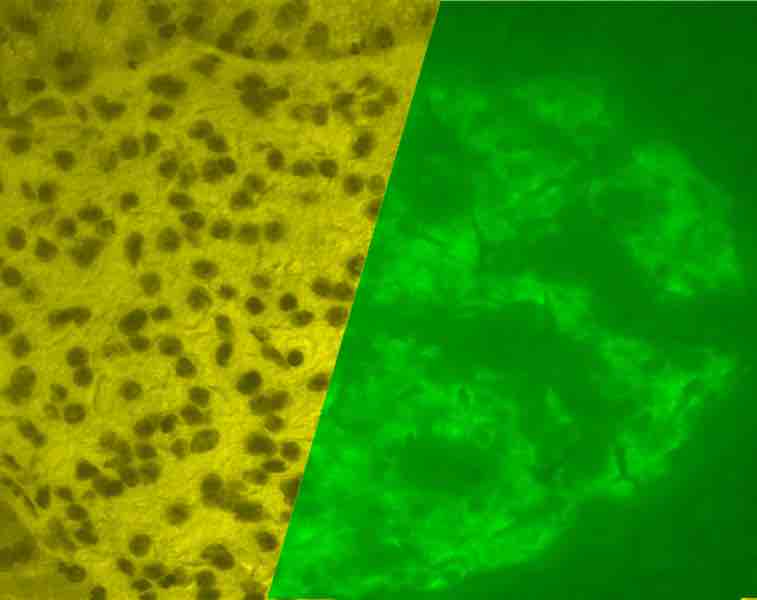
A porcine islet of Langerhans. On the left is a brightfield image created using hematoxylin stain; nuclei are dark circles and the acinar pancreatic tissue is darker than the islet tissue. The right image is the same section stained by immunofluorescence against insulin, indicating beta cells.
This is a microscope photograph of a porcine islet of Langerhans. On the left is a brightfield image created using hematoxylin stain; nuclei are dark circles and the acinar pancreatic tissue is darker than the islet tissue. The right image is the same section stained by immunofluorescence against insulin, indicating beta cells.
Source
Boundless vets and curates high-quality, openly licensed content from around the Internet. This particular resource used the following sources: