Electron Microscopy
Electron microscopy uses a beam of electrons as an energy source. An electron beam has an exceptionally short wavelength and can hit most objects in its path, increasing the resolution of the final image captured. The electron beam is designed to travel in a vacuum to limit interference by air molecules. Magnets are used to focus the electrons on the object viewed.
There are two types of electron microscopes . The more traditional form is the transmission electron microscope (TEM). To use this instrument, ultra-thin slices of microorganisms or viruses are placed on a wire grid and then stained with gold or palladium before viewing, to create contrast. The densely coated parts of the specimen deflect the electron beam and both dark and light areas show up on the image. TEM can project images in a much higher resolution—up to the atomic level of thinner objects.
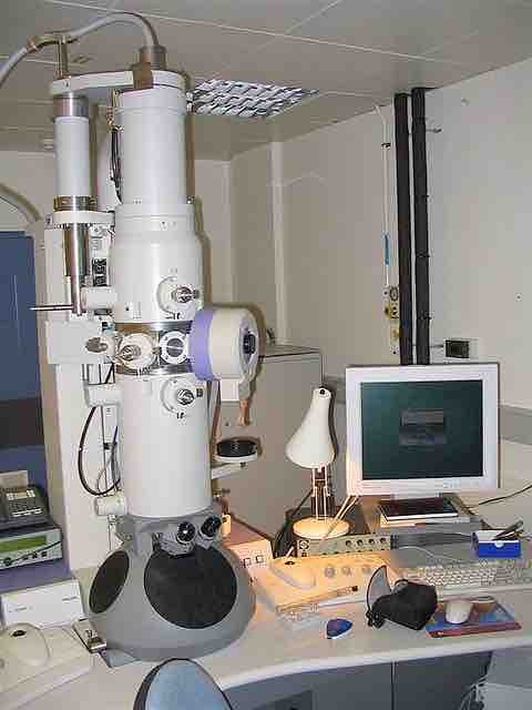
Electron microscope
A modern electron microscope
The second and most contemporary form is the scanning electron microscope (SEM). It allows the visualization of microorganisms in three dimensions as the electrons are reflected when passed over the specimen. The same gold or palladium staining is employed.
Electron microscopy has multiple applications. It is ideal to:
- explore the in vivo molecular mechanisms of disease;
- visualize the three dimensional architecture of tissues and cells;
- determine the conformation of flexible protein structures and complexes;
- observe individual viruses and macromolecular complexes in their natural biological context.
Sample preparation can be critical to generate a successful image because the choice of reagents and methods used to process a sample largely depends on the nature of the sample and the analysis required.